
VIP member
Modular algae phenotype imaging analysis system
Modular algae phenotype imaging analysis system
Product details
Modular algae phenotype imaging analysis system
 Algae are a general term for a series of aquatic organisms such as cyanobacteria, diatoms, dinoflagellates, green algae, brown algae, and red algae. There are various forms of microalgae, ranging from micrometer sized single celled microalgae to large brown algae that can grow up to several meters or even tens of meters in length. Algae, as the most important primary producers in water bodies, play an extremely important role in the stability of the entire ecosystem and even the Earth's sphere. Meanwhile, many economic algae also play important roles in industries such as food, medicine, and energy. Harmful ecological phenomena such as algal blooms and red tides are also caused by algae.
In algae research, it is necessary to comprehensively analyze the phenotypic characteristics of algae, especially the measurement and analysis of photosynthetic physiology, morphology, color, pigment composition and distribution, photosynthetic contributions of different pigments, stress physiology, etc., in order to digitize the phenotype of algae and visualize their physiological ecology and functions. This requires a phenotype imaging system specifically designed for algal phenotypes.
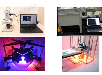
The modular algae phenotype imaging analysis system consists of FMT150 algae cultivation and online monitoring unit, FluorCam chlorophyll fluorescence or multispectral fluorescence imaging unit, FKM multispectral fluorescence dynamic microscopy imaging unit, hyperspectral imaging unit (or multispectral imaging unit), RGB color imaging unit, etc. It is currently the most flexible, comprehensive, cost-effective, and technologically advanced algae phenotype and physiological ecology research and analysis system on the market.

Phenotypic imaging study of coral symbionts, left image: NDVI multispectral imaging and GFP green fluorescent protein imaging analysis at different stages of coral contraction expansion; Right picture; Analysis of RGB imaging, chlorophyll fluorescence imaging, and NDVI multispectral imaging of coral surface and vertical profile samples under low light and strong light adaptation conditions, respectively(Leal MC,et al.2015)
Main functional features and technical indicators:
1. FMT150Algae cultivation and online monitoring technology: uniquely combining bioreactors with monitoring instruments for modular and precise light cultivation and physiological monitoring of freshwater, seawater algae, blue-green algae, etc
1)  Size and capacity options: 400ml, 1L, 3L, etc., can be customized with large culture containers of 25L, 120L, etc
2) Full LED light source: red light, blue light or dual color light source of white and red light, other light qualities can be customized, with a maximum light intensity of 3000 μ mol (photons). m-2.s-1
3) Accurately control temperature, light intensity, cultivation period CO2Cultivation conditions such as concentration, simulating natural cyclic changes, and capable of constant or constant turbidity cultivation
4) Real time online monitoring of temperature, pH value, dissolved oxygen O2/CO2Various environmental and physiological parameters such as flux, nutrient changes, optical density, chlorophyll fluorescence parameters (reflecting stress and physiological status), etc
5) Realize external control, real-time online monitoring and storage of data through dedicated computer software
6) Optional MC1000 8-channel algae cultivation and online monitoring unit for rapid and repetitive cultivation experiments

Multi omics analysis of Chlamydomonas reinhardtii, left image: precise control culture using FMT150; Right image: Chlorophyll fluorescence analysis using FluorCam (Strenkert, 2019, PNAS)
2. FluorCamChlorophyll fluorescence imaging technology: Different models and imaging sizes of chlorophyll fluorescence imaging systems can be selected according to needs, or steady-state fluorescence imaging such as GFP/YFP, PAR absorption/NDVI imaging, etc. can be selected:
1) Portable chlorophyll fluorescence imaging, with an imaging area of 31.5x41.5mm, can be used for field and laboratory chlorophyll fluorescence imaging analysis. It can also be equipped with a photosynthesis combined system for photosynthesis measurement and chlorophyll fluorescence imaging analysis measurement
2) The closed (integrated) chlorophyll fluorescence imaging system is the most comprehensive (with all protocols including OJIP, QA reoxygenation kinetics, etc.), cost-effective, and user-friendly desktop plant phenotype imaging analysis equipment. It can simultaneously image and measure plant PAR absorption, spectral reflectance index NDVI, and other parameters
3) FluorCamModular chlorophyll fluorescence imaging system, available in 13x13cm standard version and 20x20 large version, with modular structure and scalability, can be equipped with different excitation light sources such as white LED light source (used to simulate natural light source), cyan LED light source, green LED light source, red LED light source, blue LED light source for light quality experiments, used to excite different algal photosynthetic pigment proteins (the following figure shows the portable, integrated, and modular chlorophyll fluorescence imaging systems in sequence)
4) FluorCamLarge scale chlorophyll fluorescence imaging platform, with an imaging area of up to 35x35cmThe following images show portable, integrated, and modular chlorophyll fluorescence imaging systems, respectivelyLarge scale chlorophyll fluorescence imaging platform)

5) FluorCamThe multispectral fluorescence imaging technology solution is equipped with UV ultraviolet light and dedicated filters to excite and detect the multispectral fluorescence of algae, especially suitable for secondary metabolomics and disease phenotype research. There are integrated (imaging area 13x13cm) or modular (imaging area 13x13cm or 20x20cm) options available, and a large laboratory multispectral fluorescence imaging platform can also be selected

Ocean University of China uses FluorCamResearch on Multi Spectral Fluorescence Imaging System for Porphyra yezoensisPyropia yezoensisSecondary metabolic response after infection with red rot disease (Tang L, 2019)
3. FKMMultispectral fluorescence dynamic microscopy imaging technology: a microscopy imaging technology based on FluorCam chlorophyll fluorescence imaging technology. It consists of an enhanced microscope with expandable components, a high-resolution CCD camera, an excitation light source group, a spectrometer, a temperature control module, corresponding control units, and dedicated workstations and analysis software. It can not only perform chlorophyll fluorescence and multispectral fluorescence imaging analysis on microalgae, single cells, single chloroplasts, and even granule matrix thylakoid fragments, but also perform imaging analysis on fluorescent proteins, fluorescent dyes, and algal specific photosynthetic pigments.
1) Built in all programs for current chlorophyll fluorescence research, such as Fv/Fm, Kautsky induction effect, fluorescence quenching, OJIP rapid fluorescence response curve, QA re oxidation, etc., can obtain more than 70 parameters and their imaging maps
2) Equipped with 10x, 20x, 40x, 63x, and 100x specialized biological fluorescence objectives, chloroplasts and their emitted fluorescence can be clearly observed
3) The excitation light source group includes infrared light, red light, blue light, green light, white light, ultraviolet light, and far red light, which can study any pigment molecule or chromophore in plants/algae
4) Can perform imaging analysis of fluorescent proteins and dyes such as GFP, DAPI, DiBAC4, SYTOX, CTC, etc
5) High resolution spectrometers can deeply analyze various fluorescent spectra.
6) The temperature control system can ensure that the experimental samples are measured under the same temperature conditions, improve experimental accuracy, and can also be used for high/low temperature stress research.

Photosynthetic patterns during the differentiation of alien cells in Anabaena sp. (Ferimazova, 2014)
4.  SpecimHyperspectral imaging technology, recommended IQ hyperspectral imager, integrated design, built-in automatic push scan, data acquisition and processing, operating system, touch screen and operation keys, GPS, etc. The body is small and lightweight, only 1.3kg, realizing easy handheld operation or fixed operation, with a band range of 400-1000nm, 7nm spectral resolution, 204 bands, image resolution of 512x512 pixels, field of view of 31 degrees, object distance of 15cm to infinity, 1m field of view of 55x55cm. Other scanning hyperspectral imaging analysis units can be optionally configured (see the reference table for hyperspectral imaging technology selection below)
model |
FX10 |
PFD4k |
sCMOS |
FX17(nm) |
SWIR(nm) |
Band range |
400-1000nm |
950-1700 |
1000-2500 |
||
Spectral resolution (FWHM) |
5.5nm |
3.0nm |
2.9nm |
8nm |
12nm |
band |
224 |
768 |
946 |
224 |
288 |
Spatial resolution (pixels) |
1024 |
1775 |
2184 |
640 |
384 |
aperture |
F/1.7 |
F/2.4 |
F/2.4 |
F/1.7 |
F/2.0 |
signal-to-noise ratio |
600:1 |
1000:1 |
1050:1 |
||
Frame rate (fps) |
330 |
100 |
100 |
670 |
450 |
weight |
1.26kg |
2.7kg |
>2.0kg |
1.56kg |
>14kg |
5. RGB imaging analysis or multispectral imaging analysis can be selected simultaneously
6. Flexible configuration and easy to use, with the option to choose different unit combinations

Spectral imaging analysis of marine green algae, brown algae, and red algae (Ginneken V, 2017)
Online inquiry
-
Contacts
-
Company
-
Telephone
-
Email
-
WeChat
-
Verification Code
-
Message Content
-